Peculiar medical research – round 1
It is often that I come across some rather interesting medical research papers. Sometimes they are funny or strange, but always unexpected. This is why I decided to start some sort of a column where I will present these papers.
For the first time, I chose a paper (actually two papers, but they are from the same series and on the same subject) written by A. Faix, J.F. Lapray, C. Courtieu, A. Maubon and K. Lanfrey from Department of Urology and Radiology, CMC Beau Soleil, Montpellier, France.
Their titles are Magnetic Resonance Imaging of Sexual Intercourse: Initial Experience and Magnetic Resonance Imaging (MRI) of Sexual Intercourse: Second Experience in Missionary Position and Initial Experience in Posterior Position, and were both published in Journal of Sex & Marital Therapy.
Authors best describe their research in abstracts.
Magnetic Resonance Imaging of Sexual Intercourse: Initial Experience
The objective of this study was to investigate sexual intercourse with magnetic resonance imaging (MRI). A volunteer couple (30 yearold male, 27-year-old female) with a normal sex life, had face to face sexual intercourse (reversed missionary position) under MRI. Static and dynamic T2-weighted sagittal sequencies were acquired on the midline before and during vaginal penetration. In this position, before penetration, the vagina was parallel to the pubococcygeal line and had normal anterior convexity. After penetration, accentuation of the vaginal convexity was observed, produced by the penile gland reaching the anterior cul-de-sac and contact with the anterior vaginal wall. The posterior bladder wall was pushed forward and upward, the uterus upward and backward.
In this initial experience, we observed a preferential contact of the penis in erection with the anterior vaginal wall and the anterior cul-desac in the face-to-face sexual position. MRI allows a noninvasive assessment of sexual intercourse.
Magnetic Resonance Imaging (MRI) of Sexual Intercourse: Second Experience in Missionary Position and Initial Experience in Posterior Position
Our objective was to confirm that it is feasible to take images of the male and female genitals during coitus and to compare this present study with previous theories and recent radiological studies of the
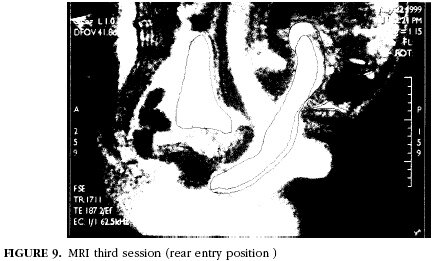
anatomy during sexual intercourse. Magnetic resonance imaging was used to study the anatomy of the male and female genitals during coitus.Three experiments were performed with one couple in two positions and after male ejaculation. The images obtained confirmed that during intercourse in the missionary position, the penis reaches the anterior fornix with preferential contact of the anterior vaginal wall. The posterior bladder wall was pushed forward and upward and the uterus was pushed upward and backward. The images obtained from the rear-entry position showed for the first time that the penis seems to reach the posterior fornix with preferential contact of the posterior vaginal wall. In this position, the bladder and uterus were pushed forward.
A different preferential contact of the penis with the female genitals was observed with each position. These images could contribute to a better understanding of the anatomy of sexual intercourse.
And now something all of you have been waiting for, the actual IMAGES.
Click to enlarge (I mean the images).
If nothing else, this volunteer couple became favorites of every “strangest places you made love” contest.
Images copyright © 2002 Brunner-Routledge
Ivor Kovic
Latest posts by Ivor Kovic (see all)
- Intraosseous access video lecture - May 22, 2015
- Emergency Physicians on Twitter - March 16, 2015
- Protected: Ddimer audit - March 12, 2015


